
Microstructure is the internal organization of a material: the size, shape, distribution, and connectivity of its particles, pores, coatings, and other features.
Techniques such as micro-CT imaging, FIB-SEM, and quantitative microstructure analysis allow scientists to visualize and measure internal architecture without destroying the sample, or at very high resolution. They connect what a material is built like to how it behaves.
A useful way to think about microstructure is to distinguish composition from organization.
Two materials can contain the same components in the same proportions and still behave differently because those components are arranged differently in space. Composition is what is present. Microstructure is how it is arranged once the product is made.
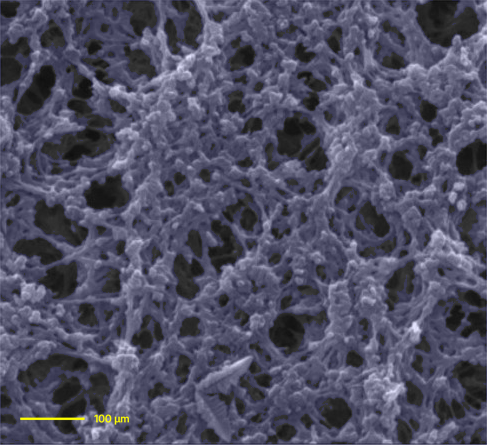
Microstructure can move performance as much as the formula does. Two dosage forms can carry the same active dose yet behave differently in use because their internal structures differ.
Release is the clearest case. Before a drug can do anything, it has to become available to the body. The rate of that release is controlled by structural factors such as particle size, packing density, porosity, and how easily fluid travels through the material to reach the drug.
This matters most in products built to release over days, weeks, or months. In long-acting injectables, implants, and topicals, microstructure is not a side detail. It is the release mechanism. A small change in internal architecture alters the rate, duration, and uniformity of release.
For this reason, developers and regulators put real weight on understanding, controlling, and measuring structure. Reliable characterization gives a team a clear view of what is actually happening inside the product.
Because many relevant features are internal, the obvious move is to section the sample and examine it directly. A focused ion beam scanning electron microscope (FIB-SEM) does exactly that at very high resolution. The tradeoff is real: the preparation destroys the structure of interest and shows only a few 2D planes. Serial FIB-SEM rebuilds a full 3D image, but it is slow and costly.
Other methods reach the inside without destroying the sample. The principle is the same as a clinical CT scan, where many projections combine to reconstruct internal structure. Here the subject may be a single particle, microsphere, coating, or porous matrix, at a far smaller scale.
Different techniques expose different structural information. Some are built for 3D volume, some for surface detail, some for the finest spatial resolution. The right one depends on the material, the feature size, and the question you are trying to answer.
Imaging is only the first step, not the finish. To compare samples, assess reproducibility, or stand behind a formal decision, the structure has to become numbers. Once measured, microstructure becomes a set of parameters you can track across batches, formulations, and process conditions.
The descriptors that carry the most weight:
Together these turn a picture into a precise description that two teams can agree on and build decisions around.
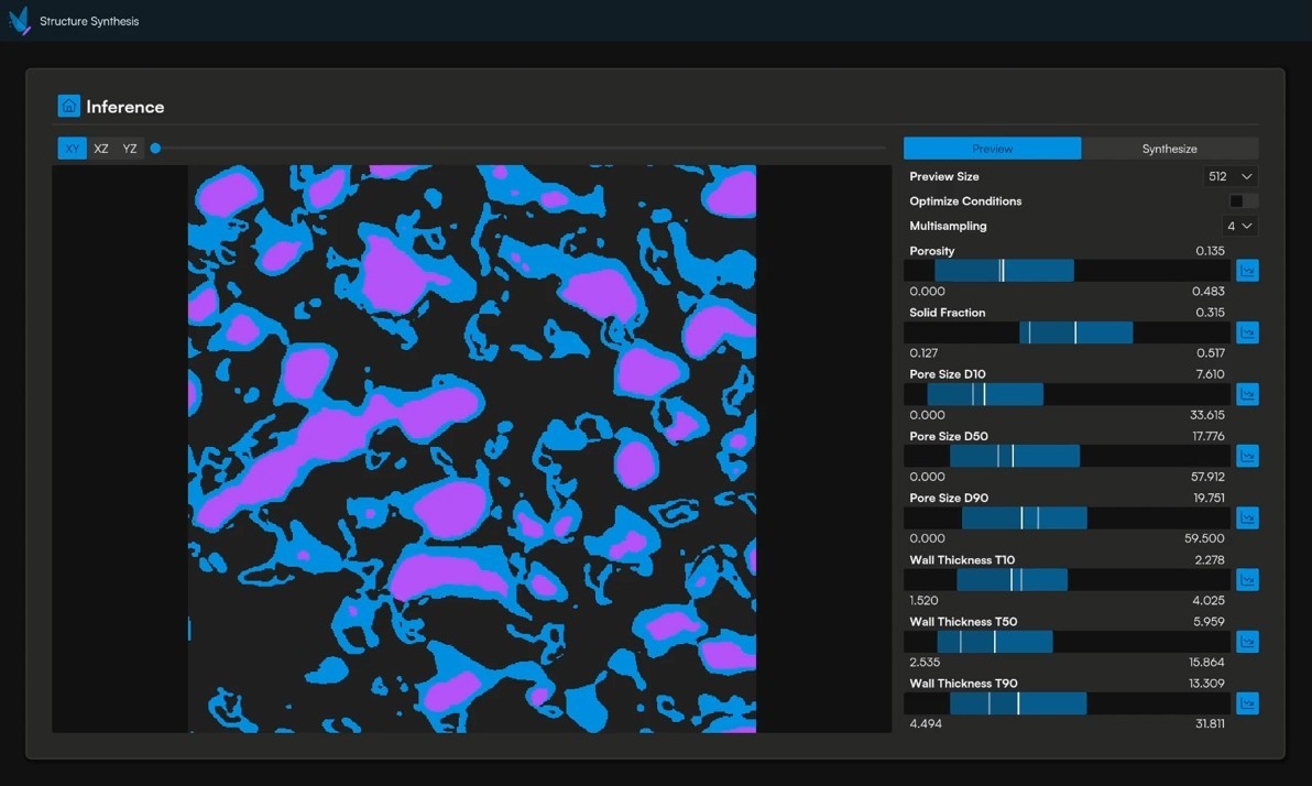
Once structure is imaged and quantified, that data feeds simulation, and simulation predicts how the material will behave before a single physical test is run.
In practice that means asking sharp questions on a computer. What does a smaller particle size do to release? More pore connectivity? A shift in where the active sits? You work through the options in simulation and only make the formulations worth making.
This progression, from visualization to quantification to prediction, is what makes microstructure analysis valuable. Imaging reveals the architecture, quantification pins it down, and modeling connects it to how the product performs.
Keep reading
Keep exploring