Image Segmentation
Fast, intuitive cloud-based segmentation for 2D pharmaceutical images; built for integration with analysis and simulation workflows.

What is Segmentation
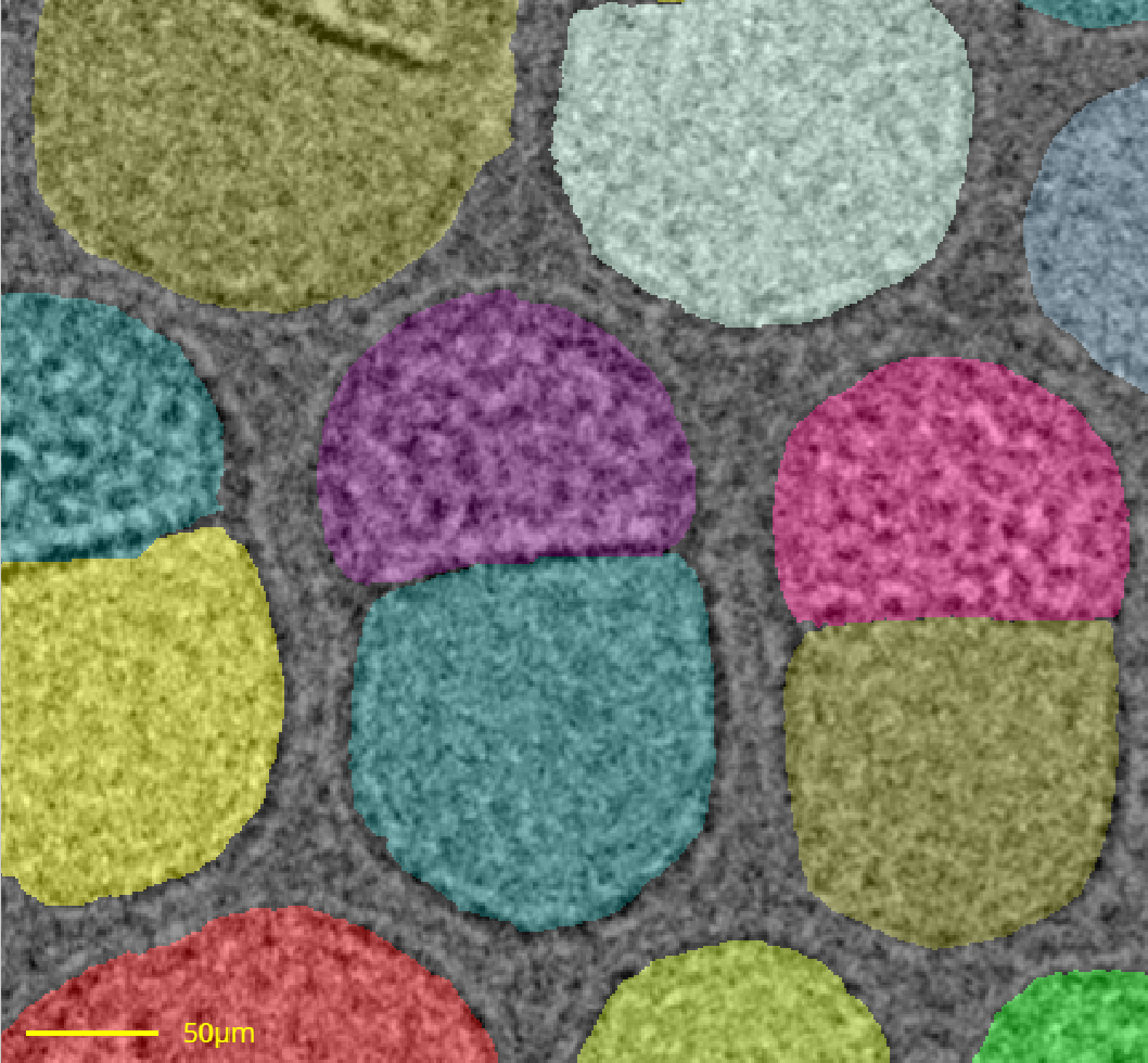
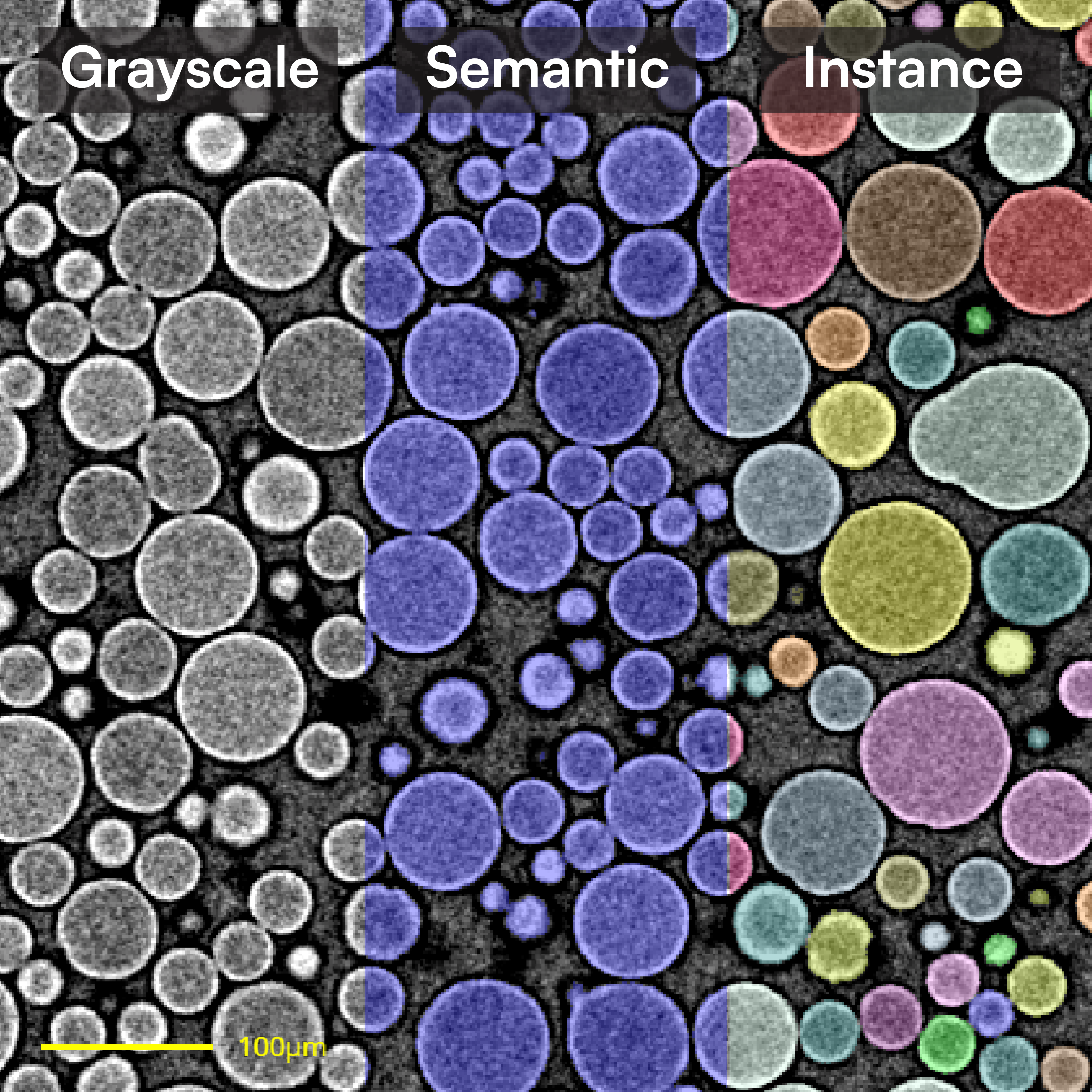
digiM I2S image segmentation tools assign pixel (2D) and voxel (3D) greyscale or color values to a feature of interests. Semantic segmentation refers to the assignments into material phases, e.g., air void space corresponding to porosity. Instance segmentation refers to further assignments into objects, e.g. individual pores from porosity phase. The computer stores digital images in greyscale or color values. It does not know which greyscale value corresponds to what material. Segmentation thus transfers human knowledge of greyscale to material correspondence into a label of that material, e.g., solid material with label of 0 and porosity with a label of 1 .
Why Segmentation is Important
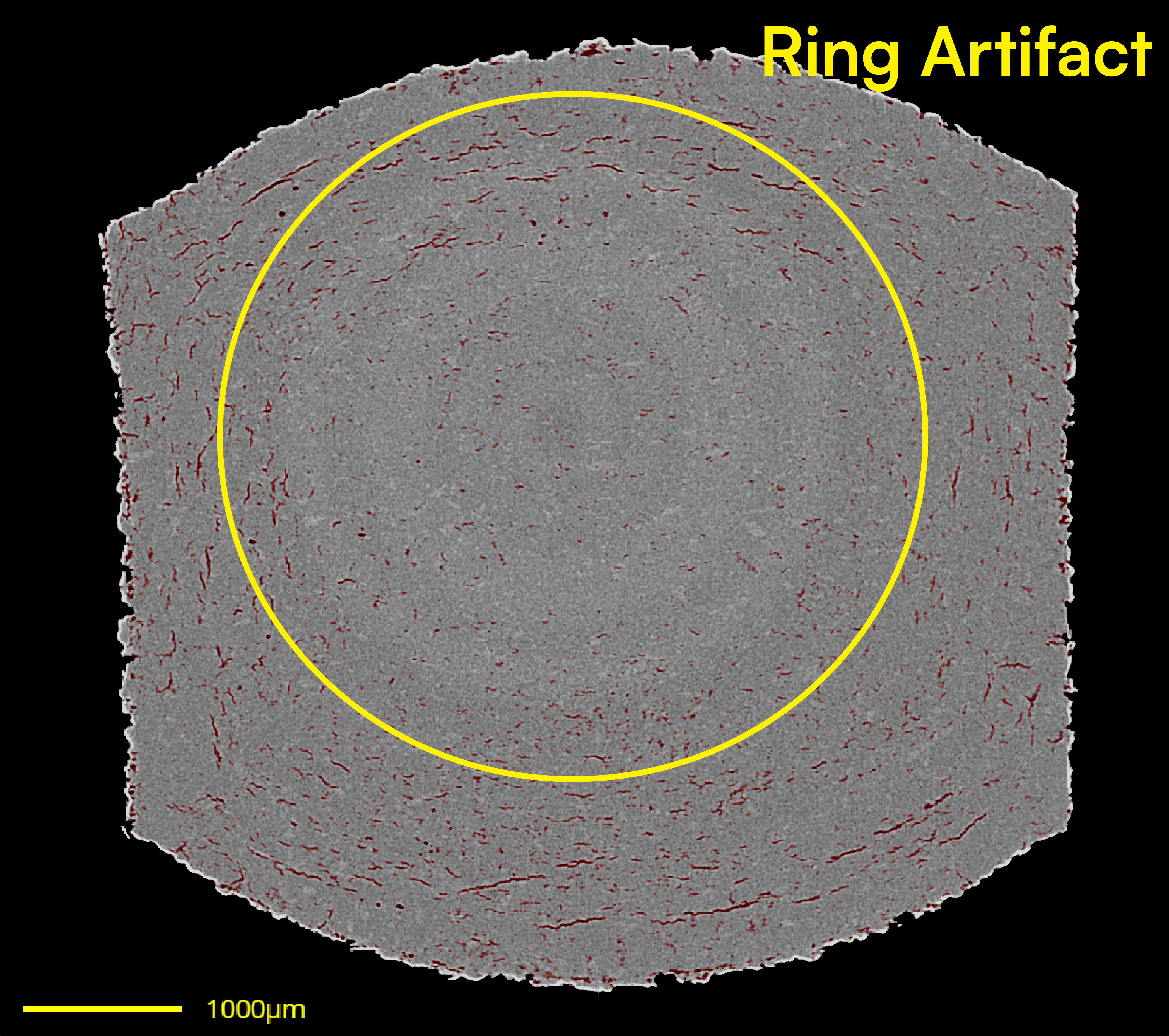
Through the labels generated by digiM I2S segmentation, material phases and objects in the images can be quantified . For example, through the segmentation of solid phase and air void phase, porosity can be calculated by counting the voxels corresponding to air void phase, divided by all voxels corresponding to both phases. Segmented voxels will further allow image-based modeling. Segmentation also allows the user to mitigate the uncertainties, artifacts, and intrinsic limitations associated with sample and imaging.


Segmentation Challenges
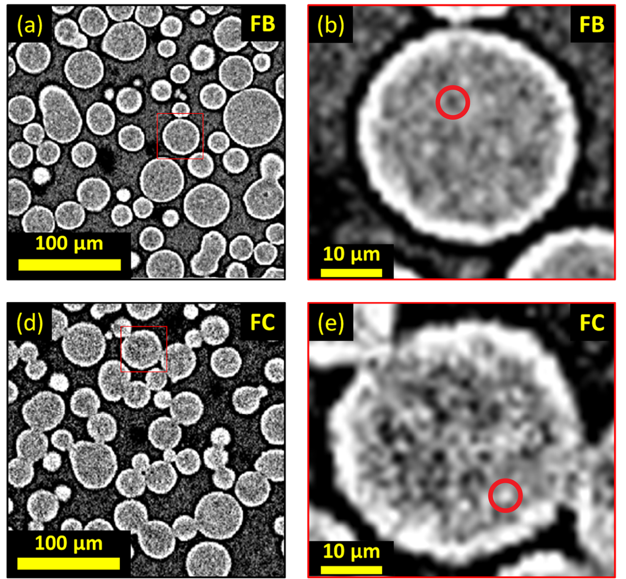
In a perfect imaging experiment, when the source signal, the interaction between the signal and the sample, and the detection, images produce sharp contrast between material phases. Simple thresholding, i.e., a cut off value of 80 in an 8-byte image with greyscale ranges between 0 and 255, will be sufficient to segment dark air void from the rest. Real world imaging experiments, however, are subjected to imperfectness compounded from all steps. The source signal is often a polychromatic wave, causing complex interactions with the sample including diffraction, deflection, and transmission. The sample might respond to the signal in an unpredictable manner, including reacting to the signal. The detectors have detection sensitivity limits and various detection artifacts . digiM I2S allows the user to execute an effective segmentation method that can achieve accurate, quality controlled, and validated segmentation in a timely manner.
Supervised Machine Learning (SML)
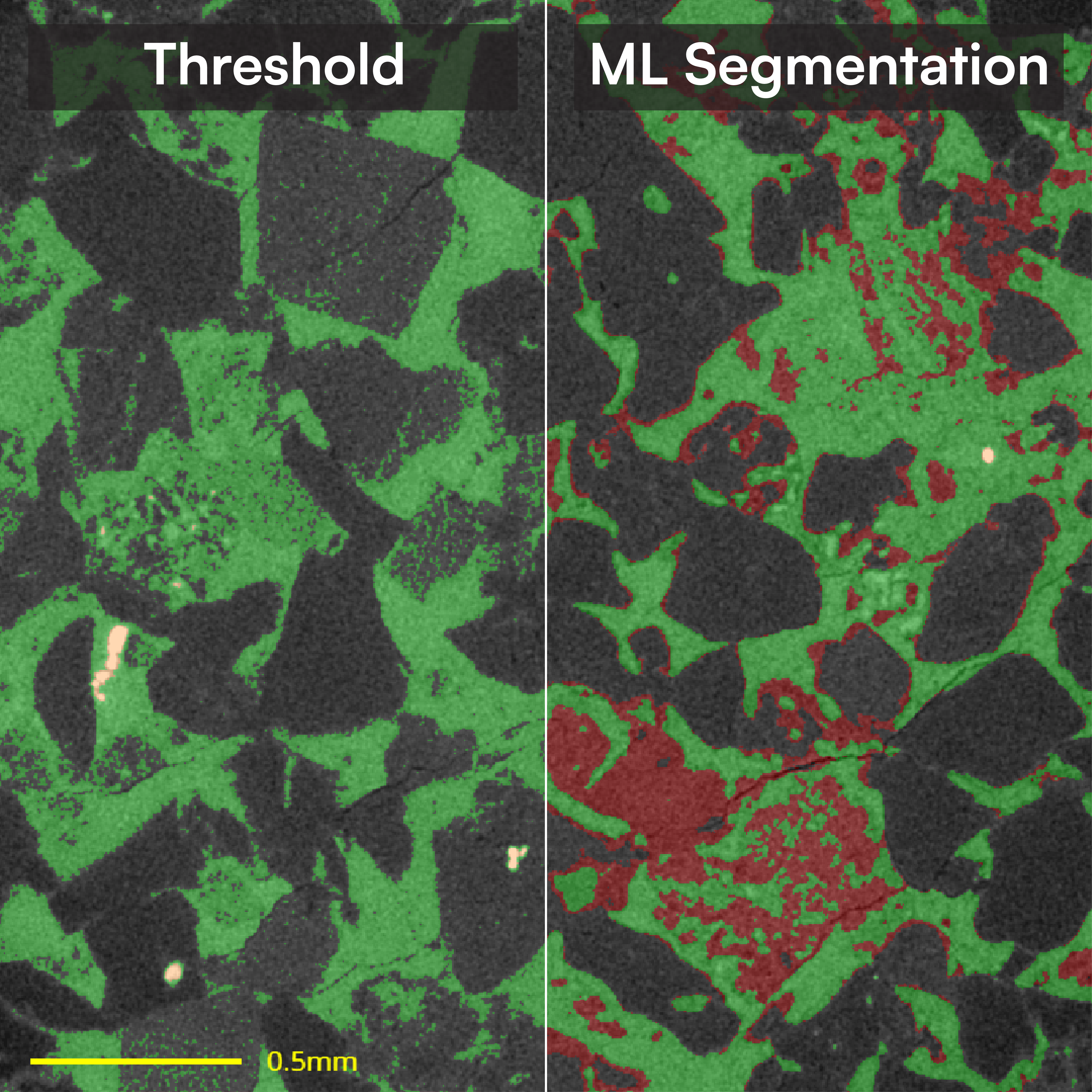
I2S machine learning segmentation uses image specific supervised training to rapidly identify the complex patterns found within pharmaceutical imaging applications. Whether working with high-noise SEM images or low-contrast micro-CT images, machine learning offers an adaptable framework to segment your imagery. I2S supervised training approach ensures accuracy by providing domain expert intervention at each step of the segmentation workflow to ensure accurate and efficient model refinement .
Deep Learning (DL)
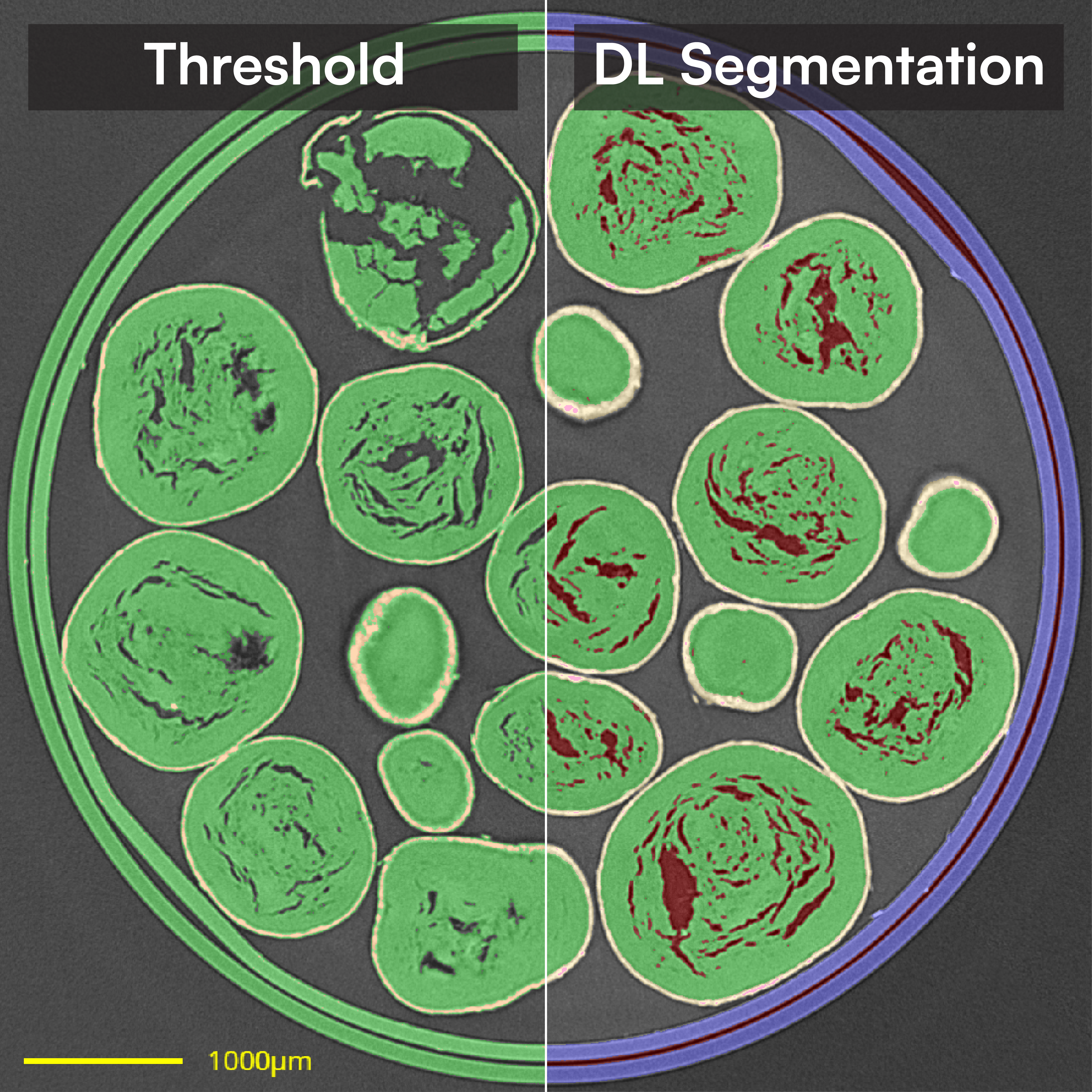
State of the art deep learning segmentation empowers I2S users to segment even the most complex images. Whether handling a few images or optimizing high-throughput workflows, I2S deep learning ensures the highest accuracy segmentation can be obtained with speeds above and beyond any other tools. Integrated with the I2S data management framework, I2S deep learning supports the management of training datasets, DL model generation, DL training refinement, and DL model reuse, all in one-place, allowing quick and easy model development, testing, and deployment .
Analysis Ready Segmentation
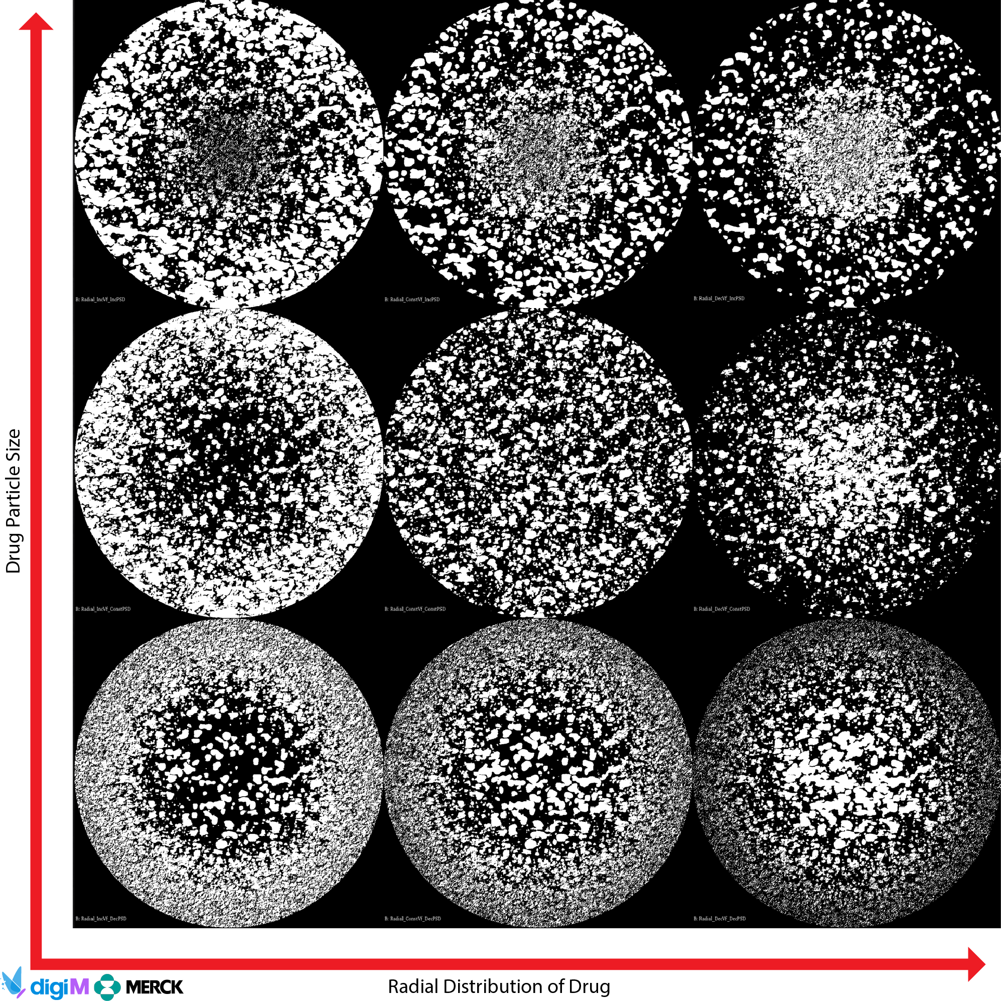
I2S SML and DL segmentations can be further refined to meet the requirements of downstream quantifications and simulations. Users can clean up segmentations, relabel particles, separate touching structures, fill voids, and more using the extensive set of editing tools. This ensures segmentations are not just visually accurate but computationally consistent for regulatory-ready analyses and simulations.

Transform Your Program with Microstructure Science
Get started with a drug product digital twin.
View Additional Software